

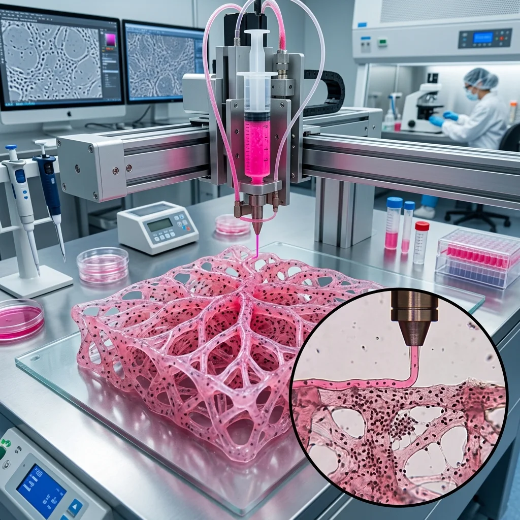
In 2025–2026, bioprinting of vascularized tissues represents the most transformative frontier in additive manufacturing: moving beyond polymers and metals to living cells, hydrogels, and growth factors printed into three-dimensional structures that mimic biological organs. The critical breakthrough is the ability to engineer vascular networks within printed tissue constructs.
Why Vascularization is Critical
In living tissues, every cell must be within < 200 μm of the nearest capillary. This boundary is set by the diffusion limit of oxygen ($O_2$) in biological media. Without an embedded vascular network, cells beyond this distance die from hypoxia within hours.
Interactive Filament Selector
Answer 3 quick questions and find the optimal material for your project.
1. What are the mechanical requirements for the part?
2. What temperature will the part operate at?
3. Are there any specific environmental requirements?
Our recommendation:
PLA filament
Ideal for easy printing, model visualization and decorative parts.
Bioprinting Technologies
| Technology | Resolution | Cell Compatibility | Applications |
|---|---|---|---|
| Extrusion Bioprinting | 100 – 500 μm | High (> 85% viability) | Skin, cartilage, bone |
| Inkjet Bioprinting | 20 – 80 μm | Moderate (70–90%) | Thin tissue layers, drug testing |
| DLP/SLA Bioprinting | 10 – 50 μm | Low–Moderate (phototoxicity) | Microfluidics, vascular networks |
| LIFT (Laser-Induced) | 10 – 30 μm | High (> 95%) | Cell positioning, complex multicellular models |
Vascularization Strategies
1. Sacrificial Networks
A network of sacrificial material (e.g., Pluronic F127, gelatin, PVA) is printed, surrounded by cell-laden hydrogel. Post-printing, the sacrificial material is dissolved, leaving open channels: artificial blood vessels.
2. Coaxial Extrusion
A coaxial nozzle simultaneously extrudes an outer layer (hydrogel + cells) and an inner layer (sacrificial or hollow core), creating tubular structures: direct vascular channels.
3. Self-Assembly
Endothelial cells (HUVEC) printed within a biomatrix spontaneously form primitive capillary networks after 7–14 days of incubation.
Pros and Cons
Pros:
- Personalized Medicine: Tissues from patient’s own cells → zero immune rejection.
- Organ Shortage Solution: Potential for printed organs instead of donor waiting lists.
- Pharmaceutical Testing: 3D tissue models replace animal testing (organ-on-a-chip).
- Regenerative Medicine: Scaffolds for bone, cartilage, and skin regeneration.
Cons:
- Technology Immaturity: Fully functional printed organs (kidney, heart) are not yet reality.
- Diffusion Limit: Vascularization remains the primary challenge.
- Regulatory Barriers: Years of clinical trials before clinical use.
- Expensive Equipment: Bioprinters and bioink materials are extremely costly.
FAQ
Bioprinting Vascularized Tissues: Living Organs From A 3D Printer Calculator
Interactive optimization calculator.
The full picture
This article is one page from The Big Book of 3D Printing: 704 illustrated pages covering every technology, material and fix in one reference.
Read it on Kindle →Engineer, author of The Big Book of 3D Printing and additive manufacturing expert